🚚 Free Worldwide Shipping on All Orders!Shop Now
1 / 30
+25
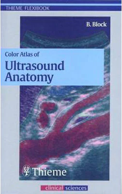
Color Atlas of Ultrasound Anatomy **
Pages: 282,
Specialty: Radiology,
Publisher: Thieme,
Publication Year: 2004,
Cover: Paperback,
Dimensions: 120x186x20mm
This brilliant pocket guide helps you to grasp the connection between three-dimensional organ systems and their two-dimensional representation in ultrasound imaging. Through dynamic illustrations and clarifying text, it allows you to: - Recognize, name, and confidently locate all organs, landmarks, and anatomical details of the abdomen -Examine all standard planes, including transverse and longitudinal scans for regions of sonographic interest (including the thyroid gland) - Understand topographic relationships of organs and structures in all three spatial planes This invaluable text is ideal for the beginner, providing a rapid orientation to all key topics. It includes: - Over 250 fully labeled image quartets, each showing: the preferred location of the transducer on the body; the resulting image; a labeled drawing of the image, keyed to anatomic structures; and a small 3-D drawing showing the location of the scanning plane in the organ. - Body markers with information on transducer handling and positioning for each sonogram - Over 250 rules of thumb and key concepts - All relevant landmarks, measurable parameters, and normal values Packed with beautiful graphics and precise text, this is the essential resource that anyone involved in ultrasound radiography needs.show more
Specialty: Radiology,
Publisher: Thieme,
Publication Year: 2004,
Cover: Paperback,
Dimensions: 120x186x20mm
This brilliant pocket guide helps you to grasp the connection between three-dimensional organ systems and their two-dimensional representation in ultrasound imaging. Through dynamic illustrations and clarifying text, it allows you to: - Recognize, name, and confidently locate all organs, landmarks, and anatomical details of the abdomen -Examine all standard planes, including transverse and longitudinal scans for regions of sonographic interest (including the thyroid gland) - Understand topographic relationships of organs and structures in all three spatial planes This invaluable text is ideal for the beginner, providing a rapid orientation to all key topics. It includes: - Over 250 fully labeled image quartets, each showing: the preferred location of the transducer on the body; the resulting image; a labeled drawing of the image, keyed to anatomic structures; and a small 3-D drawing showing the location of the scanning plane in the organ. - Body markers with information on transducer handling and positioning for each sonogram - Over 250 rules of thumb and key concepts - All relevant landmarks, measurable parameters, and normal values Packed with beautiful graphics and precise text, this is the essential resource that anyone involved in ultrasound radiography needs.show more
Pages: 282,
Specialty: Radiology,
Publisher: Thieme,
Publication Year: 2004,
Cover: Paperback,
Dimensions: 120x186x20mm
This brilliant pocket guide helps you to grasp the connection between three-dimensional organ systems and their two-dimensional representation in ultrasound imaging. Through dynamic illustrations and clarifying text, it allows you to: - Recognize, name, and confidently locate all organs, landmarks, and anatomical details of the abdomen -Examine all standard planes, including transverse and longitudinal scans for regions of sonographic interest (including the thyroid gland) - Understand topographic relationships of organs and structures in all three spatial planes This invaluable text is ideal for the beginner, providing a rapid orientation to all key topics. It includes: - Over 250 fully labeled image quartets, each showing: the preferred location of the transducer on the body; the resulting image; a labeled drawing of the image, keyed to anatomic structures; and a small 3-D drawing showing the location of the scanning plane in the organ. - Body markers with information on transducer handling and positioning for each sonogram - Over 250 rules of thumb and key concepts - All relevant landmarks, measurable parameters, and normal values Packed with beautiful graphics and precise text, this is the essential resource that anyone involved in ultrasound radiography needs.show more
Specialty: Radiology,
Publisher: Thieme,
Publication Year: 2004,
Cover: Paperback,
Dimensions: 120x186x20mm
This brilliant pocket guide helps you to grasp the connection between three-dimensional organ systems and their two-dimensional representation in ultrasound imaging. Through dynamic illustrations and clarifying text, it allows you to: - Recognize, name, and confidently locate all organs, landmarks, and anatomical details of the abdomen -Examine all standard planes, including transverse and longitudinal scans for regions of sonographic interest (including the thyroid gland) - Understand topographic relationships of organs and structures in all three spatial planes This invaluable text is ideal for the beginner, providing a rapid orientation to all key topics. It includes: - Over 250 fully labeled image quartets, each showing: the preferred location of the transducer on the body; the resulting image; a labeled drawing of the image, keyed to anatomic structures; and a small 3-D drawing showing the location of the scanning plane in the organ. - Body markers with information on transducer handling and positioning for each sonogram - Over 250 rules of thumb and key concepts - All relevant landmarks, measurable parameters, and normal values Packed with beautiful graphics and precise text, this is the essential resource that anyone involved in ultrasound radiography needs.show more
$38.25
Color Atlas of Ultrasound Anatomy **—
$38.25
Description
Pages: 282,
Specialty: Radiology,
Publisher: Thieme,
Publication Year: 2004,
Cover: Paperback,
Dimensions: 120x186x20mm
This brilliant pocket guide helps you to grasp the connection between three-dimensional organ systems and their two-dimensional representation in ultrasound imaging. Through dynamic illustrations and clarifying text, it allows you to: - Recognize, name, and confidently locate all organs, landmarks, and anatomical details of the abdomen -Examine all standard planes, including transverse and longitudinal scans for regions of sonographic interest (including the thyroid gland) - Understand topographic relationships of organs and structures in all three spatial planes This invaluable text is ideal for the beginner, providing a rapid orientation to all key topics. It includes: - Over 250 fully labeled image quartets, each showing: the preferred location of the transducer on the body; the resulting image; a labeled drawing of the image, keyed to anatomic structures; and a small 3-D drawing showing the location of the scanning plane in the organ. - Body markers with information on transducer handling and positioning for each sonogram - Over 250 rules of thumb and key concepts - All relevant landmarks, measurable parameters, and normal values Packed with beautiful graphics and precise text, this is the essential resource that anyone involved in ultrasound radiography needs.show more
Specialty: Radiology,
Publisher: Thieme,
Publication Year: 2004,
Cover: Paperback,
Dimensions: 120x186x20mm
This brilliant pocket guide helps you to grasp the connection between three-dimensional organ systems and their two-dimensional representation in ultrasound imaging. Through dynamic illustrations and clarifying text, it allows you to: - Recognize, name, and confidently locate all organs, landmarks, and anatomical details of the abdomen -Examine all standard planes, including transverse and longitudinal scans for regions of sonographic interest (including the thyroid gland) - Understand topographic relationships of organs and structures in all three spatial planes This invaluable text is ideal for the beginner, providing a rapid orientation to all key topics. It includes: - Over 250 fully labeled image quartets, each showing: the preferred location of the transducer on the body; the resulting image; a labeled drawing of the image, keyed to anatomic structures; and a small 3-D drawing showing the location of the scanning plane in the organ. - Body markers with information on transducer handling and positioning for each sonogram - Over 250 rules of thumb and key concepts - All relevant landmarks, measurable parameters, and normal values Packed with beautiful graphics and precise text, this is the essential resource that anyone involved in ultrasound radiography needs.show more